Chinese passenger airlines will be able to increase their weekly round-trip flights to the US from the current 35 to 50, starting from March 31, the US Department of Transportation (USDOT) said on Monday after frequent China-US interactions.
The approval "is a significant step forward in further normalization of the US-China market in anticipation of the Summer 2024 traffic season," the USDOT said.
The shift comes amid increased dialogue between Chinese and US officials this year.
Chinese Commerce Minister Wang Wentao met with US Trade Representative Katherine Tai on the sidelines of the 13th Ministerial Conference of the World Trade Organization in Abu Dhabi, on Monday. Wang and Tai engaged in "professional and in-depth" discussions on bilateral and multilateral economic and trade issues of mutual interest, according to the Ministry of Commerce (MOFCOM).
Wang expressed serious concerns over additional US tariffs imposed on Chinese goods and other trade issues during the talk, MOFCOM said.
In another development, the chief executive of the US Chamber of Commerce, Suzanne Clark, is leading a delegation of former US government officials to Beijing this week, Reuters reported.
The group will meet with senior Chinese government officials and local business leaders, as well as American business executives and foreign diplomats, according to a representative of the chamber.
Earlier this month Chinese and US officials held a third round of talks in Beijing as part of their Economic Working Group set up last year, adding to growing interactions since the beginning of 2024 between officials of the world's two biggest economies.
China’s Ministry of Finance (MOF) said the two sides had in-depth, candid, pragmatic and constructive exchanges on the macroeconomic situation and policies, G20 financial cooperation, debt of developing countries, industrial policies and other issues.
During the meeting, the Chinese side expressed concerns about additional US tariffs imposed on Chinese goods, restrictions against China on two-way investment, and sanctions that suppress Chinese enterprises, the MOF said, adding that the two sides agreed to continue talks.
The US delegation indicated that US Treasury Secretary Janet Yellen looked forward to a return visit to China at an "appropriate time," US Department of the Treasury said.
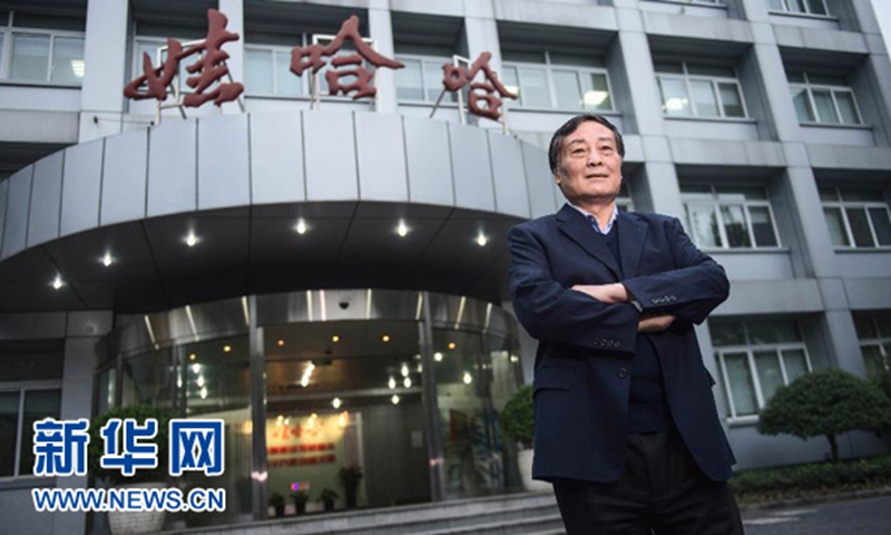
Zong Qinghou, the founder and chairman of Chinese beverage giant Wahaha Group, died on Sunday at the age of 79, the company has confirmed.
On Thursday, the company said that Zong was hospitalized but remained in stable condition. The group's business is operating as normal.
In 1987, Zong led two retired teachers to set up a school-run enterprise. With a loan of 140,000 yuan ($19,700 at current rates), they started selling soda and popsicles on a consignment basis. Later through technological and marketing innovation, they created the famous brand Wahaha.
Zong also became a major representative of China's private entrepreneurs after reform and opening-up kicked off in 1978, the Xinhua News Agency reported on Sunday.
Zong was a deputy to the 10th, 11th and 12th National People's Congresses, and a representative of the 12th, 13th and 14th National Congresses of the Communist Party of China in Zhejiang Province.
He won honors including the National Model Worker, the National May 1 Labor Medal and the "Top 100 outstanding private entrepreneurs at the 40th anniversary of reform and opening-up award."
"For Chinese entrepreneurs, it is essential to be patriotic, to innovate continuously and to care for employees," Zong once said. "Only in this way can private firms thrive."
With regard to personal wealth, Zong topped the list of the richest people in the Chinese mainland on more than one occasion. As of March 23, 2023, Zong ranked No.121 on the Hurun Global Rich List 2023 with wealth of 100 billion yuan.
As a leader in China's beverage industry, Zong created a large number of household beverage brands including Wahaha purified water and AD calcium milk. He was regarded as one of the representatives of the first generation of entrepreneurs in Zhejiang andiconic figure of China's economic reform.
Set up in 1987, the company continued to expand, with output value exceeding 100 million yuan in 1990, climbing to 10 billion yuan in 2003 and reaching 78.28 billion yuan in 2013. In 2022, the group company's sales reached 51.202 billion yuan.
Media reported that Zong had the habit of reading hard copies of major newspapers in China, which his assistants printed out for him - meaning each time he went on a business trip, his team would bring a small printer, A4 paper and ink cartridges.
Although Zong once served as the chairman of Wahaha Group, a successor has long been in place. In December 2021, Wahaha Group officially announced that Zong Fuli, daughter of Zong Qinghou, was installed as vice chairman and general manager of the group. The father did not hide his pride in her daughter, giving her 90 points in a public media interview for her performance in running the group.
In a Dialogue program of CCTV last year, Zong Qinghou addressed succession within the company. He said he would not retire, but would step back and let young people take charge of day-to-day operations.
China's Foreign Ministry said on Thursday that the model of Boeing 737 MAX8 has met delivery requirements set by Chinese regulators.
Wang Wenbin, spokesperson for the foreign ministry, said that the model is approved in accordance with Chinese civil aviation regulations on December 8 of 2023.
The comments came after media reported that Boeing delivered one Boeing 737 MAX to China Southern Airlines.
Earlier, Reuters reported that Boeing was set to deliver the first 737 MAX to a Chinese airline since March 2019 on Wednesday, citing flight data.
The delivery ends a four-year freeze for the US plane maker's most profitable jet.
China suspended most orders and deliveries of Boeing planes in 2019 after the 737 MAX model was grounded globally, following two fatal crashes.
Before the delivery, in December of last year, Boeing said that a 787-9 Dreamliner ordered by Juneyao Airlines had been delivered.
It is the first time since November 2019 that Boeing has delivered a 787 Dreamliner plane to a Chinese airline.
China's Shanghai Composite Index rose for the fourth straight day on Friday to get back above the 2,900-point mark amid the rollout of a series of policies to support the development of the capital market and the macro-economy.
Yang Delong, chief economist at Shenzhen-based First Seafront Fund Management Co, told the Global Times on Friday that the A-share market had been near the bottom level, calling for confidence and patience in the Chinese stock market.
The Central Financial Work Conference called for efforts to accelerate the building of a nation with a strong financial sector. The goal cannot be achieved without a prosperous capital market, which provides support for enterprises' transformation and upgrading as well as the country's sci-tech innovations, Yang said, adding that more policies to support the long-term and healthy development of the capital market are expected.
Pan Gongsheng, governor of the People's Bank of China (PBC), the central bank, said at a press conference on Wednesday that China will cut the reserve requirement ratio (RRR) by 50 basis points from February 5, which is expected to inject 1 trillion yuan in long-term liquidity.
Underscoring the central government's resolve to bolster the economy, the news boosted investors' confidence and reversed the stock market's recent downward trend.
On Friday, the Ministry of Commerce declared 2024 the "year of promoting consumption" and stressed the need to strengthen consumption rebound momentum. The ministry said it would continue to relax restrictions on foreign investment and improve the business environment in order to attract more investment.
Meanwhile, China's Ministry of Housing and Urban-Rural Development on Friday called on various cities to adjust real estate policies based on their local conditions, and pledged to treat developers equally in terms of financing so as to ensure the healthy and steady development of the real estate sector, China Securities Journal reported.
Investors poured almost $12 billion into Chinese equity funds in the week to Wednesday in the largest inflow seen since 2015 and the second-largest ever, Reuters reported on Friday.
The China Securities Regulatory Commission (CSRC) on Tuesday vowed to make "all efforts" to maintain the stable operation of the capital market, saying that it would put more emphasis on stability and strive to stabilize the market and investors' confidence.
An official from the CSRC also said that the country's top stock market regulators will cultivate long-term and stable investment forces, enrich policy tools to cope with market volatility and guard against risks, according to a media report on Tuesday.
These comments add to growing signals from Chinese officials that they are stepping up efforts to stabilize the capital market amid recent volatility. On Monday, China's State Council, the cabinet, called for drawing more long-term funds into the capital market to boost its inherent stability.
The signals will offer much-needed reassurance for investors that policy measures are expected to be taken to tackle risks in the capital market and ensure overall stability, which will help lift expectations and boost confidence in the capital market, analysts said on Tuesday.
A meeting of the CSRC on Tuesday stressed that the agency will make all efforts to maintain the stable operation of the capital market. It also vowed to vigorously improve the quality and investment value of listed companies and increase the entry of medium- and long-term funds, according to a statement on the commission's website.
Zhang Wangjun, a senior official at the CSRC, said that in terms of stabilizing the market and investors' confidence, the commission will enrich policy tools to cope with market volatility, hedge against risks in a timely manner and firmly hold the bottom line in preventing risks, the Xinhua News Agency reported on Tuesday.
The CSRC will also, along with relevant parties, ensure the continuity and stability of macro policies and industry policies, and avoid introducing policy measures that are not conducive to capital market expectations, in order to stabilize market expectations, according to Zhang.
The remarks from the CSRC come one day after the State Council held an executive meeting to hear reports about the capital market. The meeting stressed that increased medium- and long-term funds should be brought into the country's capital market and the inherent stability of the market should be enhanced.
The meeting also called for crackdowns on illegal activities and more effective measures to stabilize the capital market and boost investors' confidence.
Analysts said that these growing signals from government officials could mean concrete policy measures are forthcoming to tackle risks and challenges, boost investor confidence and ultimately maintain stability in the capital market, amid recent volatility.
"The recent declines in the stock market didn't happen due to major changes in the economic fundamentals, but because of irregular activities that take advantage of pessimistic narratives and market loopholes," Tian Yun, a Beijing-based economist, told the Global Times on Tuesday.
Tian said that this situation isn't in the interests of investors and will have a significant impact on the overall economy. "This is why regulators must step in to maintain financial stability by using targeted funds and closing loopholes that allow short-selling," he said.
While China mounted an impressive economic recovery in 2023, with a GDP growth rate of 5.2 percent for the year, foreign media outlets continue to push negative narratives about the Chinese economy that analysts said have had a negative impact on investors' sentiment, leading to market volatility.
As of Tuesday, the benchmark Shanghai Composite Index had fallen by 6.46 percent so far this year, while the Shenzhen Component Index had lost 8.56 percent.
Amid the downward trend, "restoring investor confidence does require strong policies and large-scale capital inflows to reverse the market's downtrend, so that investors can regain confidence," Yang Delong, chief economist at Shenzhen-based First Seafront Fund Management Co, told the Global Times on Tuesday.
The State Council meeting pointed out that it is necessary to enhance the consistency of macro policy, strengthen innovation and coordination of policy tools, consolidate and enhance the positive trend of the economic recovery and promote the stable and healthy development of the capital market.
Zhang also said that the CSRC will strengthen monitoring of the trading behavior of key investors and severely crack down on illegal transactions such as illegal trading and manipulation, according to Xinhua.
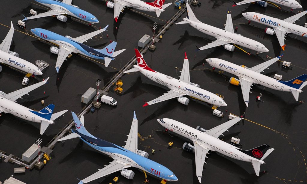
The escalating crisis of Boeing is giving rise to mounting frustration and anger within the aviation industry, as repeated safety problems of the American aviation giant, which highlight its poor quality control, along with regulatory failures and narrow-minded US industrial policies, clearly can't easily be fixed.
Two US airlines have cast doubt on Boeing 737 MAX plane orders. Scott Kirby, CEO of United Airlines, one of the biggest buyers of Boeing jets, said in an interview with CNBC on Tuesday that the MAX 9 groundings are "probably the straw that broke the camel's back," signaling to the carrier it should seek "alternative plans" for its aircraft inventory.
In another interview on NBC on Tuesday, Alaska Airlines CEO Ben Minicucci revealed the carrier found "some loose bolts on many" Boeing 737 Max 9 planes. "I'm more than frustrated and disappointed. I am angry," he said, according to excerpts of the interview.
There is every reason for airlines like United and Alaska to be disappointed. For the aviation industry, safety is the utmost bottom line, as any problem is likely to cause significant damage to life and property. Boeing's safety problems have posed a great challenge, becoming a major disruptor of the operation of airlines.
Indeed, United Airlines just warned investors that it will report a larger-than-expected loss in the first quarter because of the grounding of all 737 MAX 9 jets after a door plug blew off on an Alaska Air flight on January 5.
If anything, the CEOs' comments suggest that the safety crisis is not something that Boeing can quickly or easily quell. This is because since the crisis began earlier this month, reports of various safety problems about Boeing aircraft have continued to surface, indicating that its quality controls are facing a serious systemic problem, which cannot be easily fixed by addressing an isolated case.
For instance, a Boeing 757 aircraft lost its nose tire moments before it was supposed to take off from Hartsfield-Jackson Atlanta International Airport on Saturday, Fox Business reported on Tuesday.
Also last week, US Secretary of State Antony Blinken was forced to change planes to return to Washington from Davos after the Boeing 737 aircraft he was supposed to board suffered a critical failure related to an oxygen leak.
There has been a lot of analysis as to why Boeing is plagued by repeated mechanical mishaps, such as its over-reliance on parts outsourcing for the purpose of maximizing profits and negligence of quality control.
But these may just be superficial issues. Fundamentally speaking, Boeing's management problems that have led to defects in aircraft production are inseparable from regulatory failure and government protection.
Due to limited resources, the Federal Aviation Administration has long outsourced some of its oversight responsibilities to Boeing and other manufacturers for self-certification, according to an NPR report, meaning that Boeing has played a role as "both athlete and referee" when it comes to safety supervision.
The US government has been one of Boeing's biggest customers and its biggest supporter. It is no secret that Boeing has long been one of the largest beneficiaries of tax breaks, subsidies and loans.
When it comes to trade disputes, the US government imposed tariffs on Airbus to protect Boeing's position in the US market. Also, US officials have been pushing other countries to buy Boeing products, sometimes by offering loans.
Boeing's decline in competitiveness is due partly to the excessive support by the US government, which has made the giant lose its sense of urgency about quality controls and other issues affecting its competitive edge.
It's clear that American influence has become a more powerful guarantee of market share than production competitiveness. But such protection can backfire.
If Boeing continues to rely on US government influence around the world, rather than really addressing its safety problems, it may become the "spoiled child" of high-end manufacturing in the US and lose its pivotal position in the American manufacturing industry.
Whatever happens to Boeing, the US still leads in some manufacturing areas. As the US tries hard to bring manufacturing jobs back to the country, Boeing's crisis is worth reflecting on for the American manufacturing industry. If US industrial policy continues to be propped up by protectionism, the competitiveness of US manufacturing is bound to be cast into doubt.
Chinese Commerce Minister Wang Wentao vowed to promote healthy and stable development of China-Belgium and China-EU cooperation, and urged the EU to relax trade restrictions on high-tech products.
China is willing to expand cooperation with Europe in digital and innovative sectors, and hopes Europe can relax export restrictions on high-tech products, Wang said on Friday during a roundtable for Belgian enterprises amid the Belgian Prime Minister's visit to China.
Prime Minister of Belgium Alexander De Croo paid an official visit to China on Thursday and Friday last week, marking the first visit to China by a Belgian prime minister since 2016.
The China-Belgium and China-Europe trade structure is a reflection of multiple factors, such as the overall industrial structure, market demand and international trading conditions of the two sides.
China has never pursued a trade surplus and has expanded imports from Belgium and Europe through initiatives such as the China International Import Expo, said Wang.
Wang said that the alleged accusations of "overcapacity" related to China's new-energy sector by Europe were groundless as China's new-energy vehicle industry is making a positive contribution to the global green transition.
China firmly opposes the EU's countervailing probe into China-made electric cars, and noted that excessive government intervention in enterprises' business will bring uncertainties and risks, Wang noted.
China will actively implement the consensus reached by the leaders of the two countries, and promote cooperation between small and medium-sized enterprises in China and Belgium, said Wang.
The roundtable was the first discussion meeting hosted by China's Ministry of Commerce (MOFCOM) under the foreign enterprise roundtable mechanism, which indicates China's close attention to the economic and trade relationship with Europe, said Wang.
Representatives from over 20 Belgian enterprises appreciated China's response to their feedback on investment and cooperation in sectors such as agricultural products, biomedicine and logistics, and are willing to further facilitate China-Belgium and China-Europe economic trade development, according to MOFCOM.
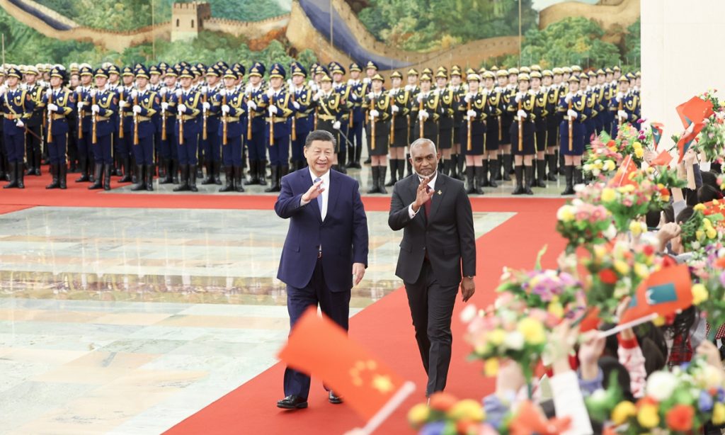
Chinese President Xi Jinping held talks with President of the Republic of Maldives Mohamed Muizzu in Beijing on Wednesday. Chinese experts said that the elevation of China-Maldives relations will bring more development opportunities to the peoples of two countries as well as to the Indian Ocean region, which is significant to China-proposed Belt and Road Initiative (BRI) and the economic and energy security of China.
According to the Xinhua News Agency, Xi welcomed Muizzi at a ceremony at the Great Hall of the People in Beijing, before holding a bilateral meeting with Muizzu, who is paying a five-day state visit to China from January 8-12. The two heads of state announced the elevation of bilateral ties to a comprehensive strategic cooperative partnership during the talks.
China stands ready to exchange governance experience with the Maldives, strengthen the synergy of development strategies, advance high-quality Belt and Road cooperation, and set a new benchmark for the China-Maldives friendship, Xi said.
He called on the two sides to strengthen cooperation in such areas as the economy, trade and investment, agricultural parks, and the blue, green and digital economies. He also called for expanded cooperation on marine ecological and environmental protection, as well as strengthened people-to-people exchanges. He said China will support more Maldivian students to study in China and promote more direct flights between the two countries.
Xi noted that the two sides should strengthen multilateral communication and coordination to safeguard genuine multilateralism and the common interests of developing countries, and build a community with a shared future for humanity to make the world more peaceful, secure and prosperous.
Muizzu said he was honored to pay his first state visit to China with a number of important cabinet ministers and become the first foreign head of state that China has hosted this year, fully demonstrating the great importance both sides attach to the development of bilateral relations.
Noting that this year marks the 10th anniversary of President Xi's historic state visit to the Maldives, Muizzu said that China has provided a significant amount of valuable assistance to his country's economic and social development. He said the Maldivian people have benefited greatly from the BRI, citing the Maldives-China Friendship Bridge as a symbol of the bond between the two peoples.
After their talks, the two heads of state witnessed the signature of an action plan to establish the China-Maldives comprehensive strategic cooperative partnership, as well as cooperation documents on the construction of the Belt and Road, disaster management, the economy and technology, infrastructure, people's livelihoods, green development, and the blue and digital economies.
Strategic significance
Zhao Gancheng, a research fellow at the Shanghai Institute for International Studies, told the Global Times on Wednesday that although Maldives is a small country in terms of land area and population, in terms of geopolitics, it has very high strategic significance.
"The Indian Ocean is far from us, but it's extremely important to the economic and energy security of our country, as well as the BRI, so China needs to try its best to make friends in the region," Zhao noted.
Xi said at the meeting with Muizzu that China respects and supports the Maldives in exploring a development path suited to its national conditions, and supports the Maldives firmly in safeguarding its national sovereignty, independence, territorial integrity and national dignity.
Muizzu said the Maldives pursues the one-China policy firmly. Firm mutual support in safeguarding national sovereignty, independence and territorial integrity is a solid foundation for the sustained and sound development of Maldives-China relations.
The Maldives supports the Global Development Initiative, the Global Security Initiative and the Global Civilization Initiative, all of which were put forward by President Xi, and is willing to communicate and cooperate closely with China on international and regional affairs, Muizzu noted.
Hu Zhiyong, a research fellow at the Institute of International Relations at the Shanghai Academy of Social Sciences, told the Global Times on Wednesday that Muizzu's visit to China shows that Maldives sincerely wants to develop ties with China and strengthen existing cooperation.
"Muizzu's pledge to the Maldivian people that there will be 'no foreign military presence,' makes India angry, so the newly elected president will surely hope China can help his country deal with the pressure from New Delhi," Hu said.
China-Maldives relations do not target any third party, but due to India's aggressive stance against its neighbor, countries in the region will definitely want to diversify their ties to preserve their national dignity and sovereignty via cooperation with other major powers like China, said Chinese experts.
This is why the normal development of ties between China and Maldives is making some India elites and decision-makers anxious, although it is unnecessary and exposes New Delhi's intention to suppress its neighbors, said analysts.
Cooperation between China and Maldives, as well as other countries in the Indian Ocean will face challenges and uncertainties from India's interruption, so China and Maldives need to work together to strengthen their security cooperation to ensure the implementation and operation of other projects, and to jointly oppose foreign intervention, Hu noted.
According to Indian media, some Indian internet users are launching a boycott campaign against Maldives amid tensions between Maldives and India, and reports claim that Muizzu is trying to attract more Chinese tourists to the Maldives to offset the loss caused by India's boycott.
Qian Feng, director of the research department at the National Strategy Institute at Tsinghua University, told the Global Times on Wednesday that Chinese tourists will return to Maldives step by step in the post-pandemic era, because it is a well-known paradise for tourism among Chinese public, so the return of Chinese tourists will not be driven by so-called political reasons or any diplomatic activity.
China had remained as the largest source of tourist arrivals in the Maldives for years before the outbreak of the COVID-19 pandemic. In 2019, nearly 300,000 Chinese tourists visited the Maldives, accounting for around 17 percent of total tourist arrivals that year, Xinhua reported.
It would be short-sighted and narrow-minded to understand the development of China-Maldives ties only based on tourism. Hu said that apart from the tourist industry, both sides can explore cooperation in other fields related to tourism, such as protection of coastal and marine ecosystems, and the development of the maritime economy, as well as public health cooperation like promoting traditional Chinese medicine to help local people to get better healthcare.